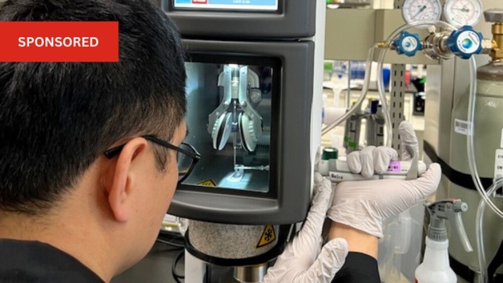
Helix Biostructures offers a full gene-to-structure suite of cryo-EM services that focuses on both quality and fast turnaround/throughput.
Over the past decade, electron cryo-microscopy (cryo-EM) has become a critical tool for structural biology. Single-particle cryo-EM has opened up high-resolution structure determination for a wide range of macromolecules that would previously have been intractable to structure solution by X-ray crystallography. Over the past 10 years, advancements in sample supports, microscope, and camera hardware, and data processing software have brought cryo-EM to a point where structures in the 2 to 3-angstrom resolution range can be generated routinely from amenable samples. In this resolution range, cryo-EM can reveal key details like small molecule binding interactions, precise epitope mapping, and details of protein:protein interactions.
Helix Biostructures offers a full gene-to-structure suite of cryo-EM services that focuses on both quality and fast turnaround/throughput. Because our lab is also equipped for protein expression and purification, we are one of the few CROs that can go from gene to vitrified cryo-EM samples without any freeze/thaw cycles or polish and vitrify pre-existing samples without added cycles. Using our milestone-based offerings, let us use our expertise to guide you through the entire cryo-EM process (including grid screening, pilot feasibility data, and high-resolution structure determination), keeping you informed with weekly updates and detailed technical reports after each milestone. With our emphasis on quality, our prices include as much data processing as needed to maximize the resolution of the final map(s) from the collected data, as well as atomic models that are thoroughly manually inspected and refined. We are proud of our record for producing excellent quality structures in high-impact publications in collaboration with our customers1-4. To date, all gene- or protein-to-structure projects delivered from our milestone-based services have achieved resolutions of 3.2 Å or better. A la carte services for any step of the cryo-EM structure determination workflow are also available.
At Helix, we approach access to microscopy hardware by following the same “synchrotron model” as our X-ray crystallography service. Sample preparation and vitrification are handled in-house by Helix scientists, but rather than owning and maintaining several multi-million-dollar microscopes and associated hardware, we purchase blocks of microscope time from facilities across the United States. This distributed network of microscopes means that we are resilient to downtime on any one instrument and can operate with much lower costs – passing those savings on to our customers. Just as with synchrotron X-ray sources, Helix scientists either carry out data collection remotely or work closely with on-site facility scientists to set the details of data collection, and we maintain full rights to the resulting data, which is securely transferred to our computational infrastructure after collection.
Transforming cryo-EM image data into high-resolution 3D potential maps requires significant computational power. Although preliminary maps can be generated in nearly real-time with data collection (given sufficient compute power and prior knowledge of the target particle), extracting the highest resolution and most clearly interpretable map(s) from a dataset requires sustained compute power over time frames that are often longer than what was spent preparing and imaging a sample. To provide fully scalable capacity for this process at Helix, all of our cryo-EM data processing is performed using Amazon Web Services (AWS) cloud computing. This means that our computational throughput is not limited by project load: Helix scientists and projects do not compete for compute resources, and our costs do not have to account for computing hardware maintenance and upgrades.
A decade has passed since the onset of the “resolution revolution” in single-particle cryo-EM. Nevertheless, there are several active areas of development working on addressing the challenges that still exist in today’s state of the art, such as: Sample supports that lead to reductions in beam-induced movement; improved phase plates that promise to allow imaging closer to focus while preserving contrast; better knowledge- and neural network-based priors for regularization during particle alignment; and use of neural networks to better model conformational variability. At Helix, we look forward to integrating these advancements into our service as they become available.
- Padi SKR, Vos MR, Godek RJ, Fuller JR, Kruse T, Hein JB, Nilsson J, Kelker MS, Page R, Peti W. Cryo-EM structures of PP2A:B55-FAM122A and PP2A:B55-ARPP19. Nature (2024).
- Bedian V, Biris N, Omer C, Chung JK, Fuller J, Dagher R, Chandran S, Harwin P, Kiselak T, Violin J, Nichols A, Bista P. STAR-0215 is a Novel, Long-Acting Monoclonal Antibody Inhibitor of Plasma Kallikrein for the Potential Treatment of Hereditary Angioedema. J Pharmacol Exp Ther (2023).
- Kwon JJ, Hajian B, Bian Y, Young LC, Amor AJ, Fuller JR, Fraley CV, Sykes AM, So J, Pan J, Baker L, Lee SJ, Wheeler DB, Mayhew DL, Persky NS, Yang X, Root DE, Barsotti AM, Stamford AW, Perry CK, Burgin A, McCormick F, Lemke CT, Hahn WC, Aguirre AJ. Structure-function analysis of the SHOC2-MRAS-PP1C holophosphatase complex. Nature (2022).
- Garvie CW, Wu X, Papanastasiou M, Lee S, Fuller J, Schnitzler GR, Horner SW, Baker A, Zhang T, Mullahoo JP, Westlake L, Hoyt SH, Toetzl M, Ranaghan MJ, de Waal L, McGaunn J, Kaplan B, Piccioni F, Yang X, Lange M, Tersteegen A, Raymond D, Lewis TA, Carr SA, Cherniack AD, Lemke CT, Meyerson M, Greulich H. Structure of PDE3A-SLFN12 complex reveals requirements for activation of SLFN12 RNase. Nature Communications (2021).


