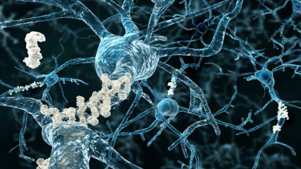
One of the common research approaches to treating brain diseases such as Parkinson’s disease (PD) and Alzheimer’s disease (AD) is using antibodies designed to clear the accumulated misfolded proteins implicated in the diseases. To date, these drug trials have not been particularly successful at improving the disease.
One of the common research approaches to treating brain diseases such as Parkinson’s disease (PD) and Alzheimer’s disease (AD) is using antibodies designed to clear the accumulated misfolded proteins implicated in the diseases. To date, these drug trials have not been particularly successful at improving the disease, even when they are effective at removing the proteins. A new theory hypothesizes that the antibodies are actually increasing the neuroinflammation associated with PD and AD, basically making the disease worse while removing the proteins.
Stuart Lipton, a researcher at Scripps Research Institute, recently published research suggesting that this may be exactly what is happening. They ran a series of in vitro assays and experiments in mice that had brain grafts of human-induced pluripotent stem cell (hiPSC)-derived microglia (hiMG). They found that the antibodies that target the misfolded proteins found in PD and AD trigger the NLRP3 inflammasome, which can lead to cell death.
“Our findings provide a possible explanation for why antibody treatments have not yet succeeded against neurodegenerative diseases,” said co-senior author Lipton, Step Family Foundation endowed chair in the department of molecular medicine and founding co-director of the Neurodegeneration New Medicines Center at Scripps Research.
The results were published in Proceedings of the National Academy of Sciences (PNAS).
The research started when Dorit Trudler, a postdoctorate researcher in Lipton’s lab, attempted to make microglia in the lab. Microglia are the innate immune cells in the brain, and it is a notoriously difficult task because the cells don’t originate from the same type of stem cells in the bone marrow that generates the rest of the immune system.
Instead of coming from the bone marrow like B and T cells and macrophages, microglia are created from the yolk sac that embryos swim in during early development, then migrate from the sac to the brain. Trudler was able to turn human-derived stem cells to turn into a yolk sac-like structure, and from there, developed cells that were identical to microglia removed from humans based on the mRNA they expressed.
Lipton indicated, “They match as closely as possible.”
They then exposed these microglia to either alpha-synuclein, the misfolded protein identified in Parkinson’s patients, and the microglia shot off inflammatory signals. And when they exposed the microglia to amyloid-beta, the hallmark of Alzheimer’s, the inflammation grew worse.
They then worked with biopharma companies to obtain antibodies that bind to either alpha-synuclein or amyloid-beta. Although Lipton is not saying which companies, it did say one of them was not Biogen’s aducanumab, which is up for approval or rejection for Alzheimer’s by June 7 by the U.S. Food and Drug Administration (FDA).
The surprising and potentially extremely important result was that although the antibodies successfully attached to their protein targets, it didn’t help with inflammation. “Rather than make things better, it actually made things worse,” Lipton said.
And they further found in humanized mice with both human and mice microglia, that the pro-inflammatory response was unique to the human cells. This means that biopharma companies, in working with laboratory animals on therapeutic antibodies for these diseases, would not have seen this reaction. Although unclear yet why it’s causing inflammation, they are convinced the NLRP3 pathway is involved, although they don’t have the exact mechanism worked out. Potentially, however, it might be possible to treat patients with a combination of the protein-clearing drugs and anti-inflammatories that block the NLRP3 pathway.
In an unrelated study, researchers at Massachusetts General Hospital utilized whole-genome sequencing (WGS) to identify rare genomic variants associated with AD. In doing so, they found 13 mutations, which establish new genetic links between AD and synaptic function. They believe it could help guide the development of new drugs for AD.
The first group of genes identified that were associated with AD involve the accumulation of amyloid-beta. The next 30 AD gene mutations identified were linked to chronic inflammation in the brain. Loss of synapses is the neurological change most closely linked with the severity of dementia in AD, but until now clear genetic association had not been identified.
Rudolph Tanzi, vice chair of Neurology and director of the hospital’s Genetics and Aging Research Unit, said, “It was always kind of surprising that whole-genome screens had not identified Alzheimer’s genes that are directly involved with synapses and neuroplasticity.”
Tanzi went on to note that identifying less-common mutations that increase the risk for AD may provide critical information about the disease. “Rare gene variants are the dark matter of the human genome,” he said.
There are many of them, as it turns out. Of the three billion pairs of nucleotide bases in the human genome, about 5o to 60 million are gene variants and 77% are rare.


