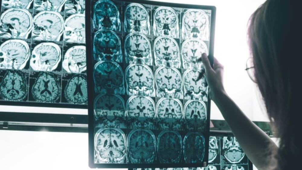
Eli Lilly and Company and its wholly-owned subsidiary Avid Radiopharmaceuticals, announced the successful Phase III trial of a Positron Emission Tomography (PET) imaging agent, flortaucipr F 18, in identifying tau in the brains of late-stage Alzheimer’s patients.
There are two primary mechanisms believed to cause the loss of memory and cognition in Alzheimer’s disease. The first is the accumulation of beta-amyloid in the brain. This occurs early on in the disease and is the focus of much of the research into preventing and treating the disease. The second is the accumulation of another type of protein tangles called tau. This tends to occur later on in the disease.
Eli Lilly and Company and its wholly-owned subsidiary Avid Radiopharmaceuticals, announced the successful Phase III trial of a Positron Emission Tomography (PET) imaging agent, flortaucipr F 18, in identifying tau in the brains of late-stage Alzheimer’s patients.
PET scans are imaging tests that utilize special dyes containing radioactive tracers. The tracers are swallowed, inhaled or injected into a vein, depending on the part of the body being studied. Specific organs or tissues absorb the tracer. The tracer gathers in areas of higher chemical activity. PET scans are typically used to measure blood flow, oxygen use, the body’s sugar metabolism and other active functions.
In the Lilly and Avid study, dubbed A16, 156 end-of-life patients with dementia, mild cognitive impairment or normal brain function received flortaucipir PET imaging. Then, 67 of the patients were studied postmortem. The trial met pre-specified endpoints with flortaucipir showing statistically significant sensitivity and specificity for identifying tau pathology of Braak Stage V/VI. This is a pathological staging scale for tau neurofibrillary tangles.
The imaging agent also demonstrated statistically significant sensitivity and specificity for total Alzheimer’s disease neuropathologic change, which combined both tau and amyloid plaque densities. This was based on the National Institute on Aging and Alzheimer’s Association (NIA-AA) neuropathology criteria.
Lilly and Avid plan to release more data in October at the Clinical Trials on Alzheimer’s Disease meeting held in Barcelona, Spain. They are in discussions with the U.S. Food and Drug Administration (FDA) about the next steps for regulatory approval.
“These encouraging results are a major advance in our ability to image the pathology of Alzheimer’s disease,” said Mark Mintun, vice president of Lilly’s pain and neurodegeneration research and development, in a statement. “We hope this and other advances in the field can help speed development of treatments, as well as provide more diagnostic information for doctors taking care of patients suspected of having Alzheimer’s.”
Alzheimer’s disease and most other forms of dementia are diagnosed through clinical symptoms, for example, forgetfulness and confusion. There are, however, several types of dementia besides Alzheimer’s disease, such as Lewy body dementia, frontotemporal dementia, and vascular dementia. Alzheimer’s accounts for 60 to 80 percent of dementias.
There have been some advances in using biomarkers, such as the genetic marker ApoE, and imaging studies. However, ApoE doesn’t diagnose Alzheimer’s, but is sometimes linked to higher risk of the disease. This PET scan imaging modality appears to be one more tool to be used in investigating Alzheimer’s and other dementias, but not necessarily for diagnosing it. It may also be useful for tracking the progression of the disease.
Interestingly, in 2017, the findings of a four-year study of PET scans in the brains of 4,000 Medicare patients with mild cognitive impairment (MCI) or dementia, was presented at the Alzheimer’s Association International Conference in London, reported The Washington Post. The study intends to evaluate more than 18,000 people, but the results were presented on the first 4,000. The Post wrote, “Among 4,000 people tested so far in the Imaging Dementia-Evidence for Amyloid Scanning (IDEAS) study, researchers from the Memory and Aging Center at the University of California at San Francisco found that just 54.3 percent of MCI patients and 70.5 percent of dementia patients had the plaques. A positive test for amyloid does not mean someone has Alzheimer’s, though its presence precedes the disease and increases the risk of progression. But a negative test definitively means a person does not have it.”


