Researchers from the Keck School of Medicine of the University of Southern California developed a new nanoparticle technique that “lights up” the unstable calcifications (plaque) that can cause heart attacks and strokes.
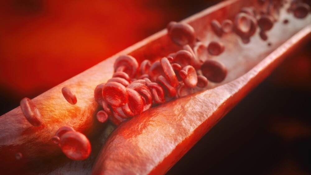
Atherosclerosis is caused by the build-up of fatty deposits, inflammation and plaque on blood vessel walls. It is estimated that by the age of 40, about half of the population has the condition, often without symptoms. However, it is a key underlying factor in heart disease and stroke.
The plaque deposits are made up of cholesterol, fatty substances, cellular waste products, calcium and fibrin, a clotting factor in the blood. When the plaque builds up, the blood vessel walls thicken, which decreases blood flow. This results in lower levels of oxygen and other nutrients reaching tissues. These plaques can develop anywhere in the body. When they occur in the arteries in or leading to the heart, it is called coronary heart disease. When it occurs in the extremities, it is peripheral artery disease.
If a piece of the plaque breaks off, it can be carried in the blood until it is lodged somewhere, which can result in a blood clot. If this occurs in an artery supplying blood to the heart or brain, it can result in a heart attack or stroke.
Researchers from the Keck School of Medicine of the University of Southern California (USC) developed a new nanoparticle technique that “lights up” the unstable calcifications (plaque) that can cause heart attacks and strokes. They published their research in the Royal Society of Chemistry’s Journal of Materials Chemistry B.
“An artery doesn’t need to be 80% blocked to be dangerous,” said Eun Ji Chung, the Dr. Karl Jacob Jr. and Karl Jacob III Early-Career Chair, senior author of the study. “Just because it’s a big plaque doesn’t necessarily mean it’s an unstable plaque.”
The research was conducted by PhD student Deborah Chin under the supervision of Chung in collaboration with Gregory Magee, assistant professor of clinical surgery from Keck.
When microcalcifications form within arterial plaques, they are more likely to rupture. Identifying whether that calcification is unstable is difficult with current CT and MRI scanning techniques, or angiography.
“Angiography requires the use of catheters that are invasive and have inherent risks of tissue damage,” said Chin, lead author of the study. “CT scans on the other hand, involve ionizing radiation which can cause other detrimental effects to tissue.”
Traditional imaging techniques also do not usually have the resolution to identify the smaller blockages that are prone to rupture.
The research group engineered a nanoparticle they called a micelle. The nanoparticle attaches itself to calcification and lights up. They specifically target hydroxyapatite, a form of calcium seen in arteries and atherosclerotic plaques.
“Our micelle nanoparticles demonstrate minimal toxicity to cells and tissue and are highly specific to hydroxyapatite calcifications,” Chin said. “Thus, this minimizes the uncertainty in identifying harmful vascular calcifications.”
So far, the technique has been tested in Petri dishes, mice, and patient-derived artery samples. The next step is to test it in targeted drug therapy to treat calcification in arteries, not just as a way to detect the potential blockages.
“The idea behind nanoparticles and nanomedicine is that it can be a carrier like the Amazon carrier system, shuttling drugs right to a specific address or location in the body, and not to places that you don’t want it to go to,” Chung said. “Hopefully that can allow for lower dosages, but high efficacy at the disease site without hurting normal cells and organ processes.”


